Hepatomegaly ultrasound 139352-Hepatomegaly ultrasound images
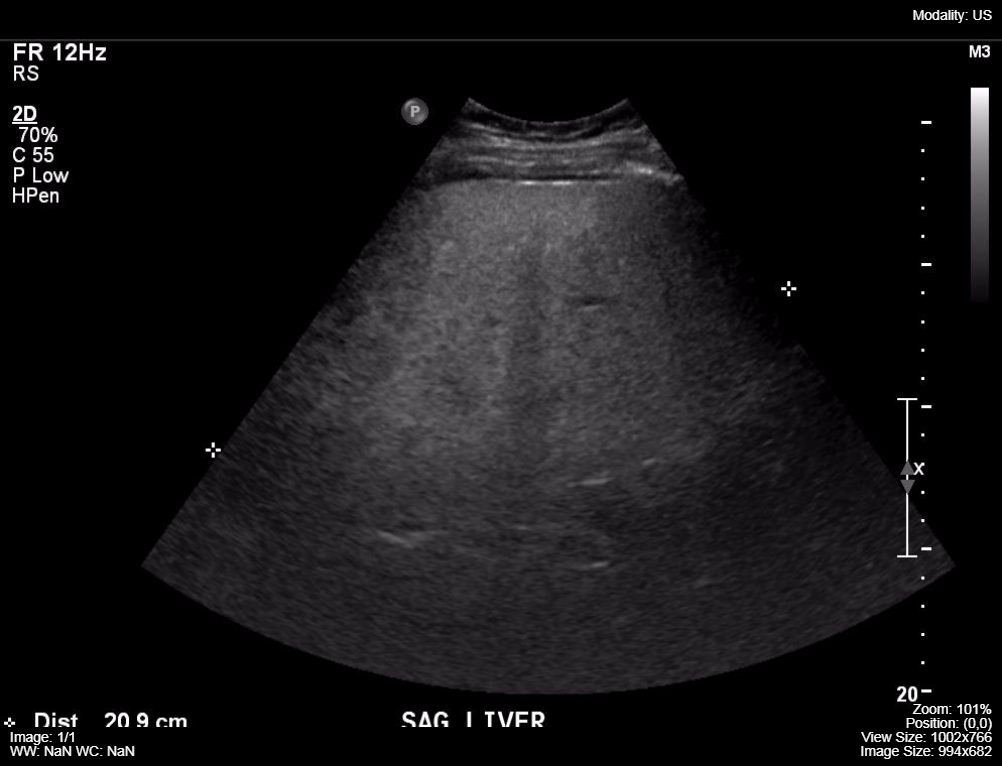
Figure 5 Longitudinal ultrasound scan of the upper fetal abdomen, demonstrating the marked enlarged fetal liver (L) and spleen (S) Day 1 Day 10 Day 21 White blood cells (gpt/l) Platelet count (gpt/l) Blast forms Hematocrit 185 542 (58%) 052 78 1269 (43%) 041 8 526 (7%) 023 gpt, gigaparticle Table 1 Postnatal hematological values Hepatomegaly is the medical term for an enlarged liver It is a symptom of disease rather than a disease in itself Sometimes, hepatomegaly may be Ultrasound indicates liver is 211 cm I am overweight female Diagnosed with fatty liver and hepatomegaly Dr advised to lose weight and avoid alcohol and follow up on 3 months I have lost 14lbs, no liquor but have had 12 glasses of red wine per week ALT was 58 and AST 50 All others were within range

Dedicated To The Mission Of Bringing Free Or Low Cost Educational Materials And Information To The Global Ultrasound Community
Hepatomegaly ultrasound images
Hepatomegaly ultrasound images-Ultrasound is the initial imaging modality almost always employed to diagnose this infantile hepatic tumor Hemangioendotheliomas have a varied appearance ranging from a solitary solidappearing lesion to a multifocal variegated cystic mass Ultrasound can accurately arrive at a diagnosis if the vascular cystic component is the predominant featureHepatomegaly R160 Polycystic disease R8 SplenomegalyR161 Vascular Abdominal (Abdominal duplex/Doppler) Liver transplant Portal HTN K766 Portal venous thrombosis I90 TIPS Z958 Kidneys (Renal Ultrasound) Renal disease (CKD) N2 Polycystic kidneys Q613 Renal cyst / mass N281/N28 UT Flank / back pain R109/M549


Animal Liver Disease Long Beach Animal Hospital
Ultrasound Early in the course of the disease, the main abnormality is enlargement of the right hepatic lobe Normally the right hepatic vein measuresLiver disease Hepatomegaly is a term used to describe a liver that is enlarged beyond its normal dimensions, and ultrasound is often a front line investigation in the suspicion of hepatomegaly This study sought to develop a reference range for the size of the normal liver using a simple, reliable and valid measurement technique Ultrasound (US) echogenic liver parenchyma The hepatic veins adjacent to the vena cava are often enlarged Hepatomegaly and lymphatic periportal edema Progressive gallbladder wall thickening or small amounts of clear fluid in the perihepatic space and in the gallbladder fossa Frequently enlarged lymph nodes in the hepatic ilium
Dr Mark Hoepfner answered 39 years experience General Surgery A There are several causes of mild hepatomegaly Common among them include fatty liver, viral hepatitis, drugs, alcohol etc However, mild hepatomegaly can be a normal finding in children There may not be any symptoms related to mild hepatomegaly Sometimes, patients complain of discomfort in right upper abdomenPartial hepatomegaly can be diagnosed by ultrasound The disease has a characteristic echoprism a violation of the homogeneity of the tissues of the organ The presence of tumors, cysts or abscesses also indicate a partial change in the liver and the progression of
The most common presentation is hepatomegaly The ultrasound appearance of multiple cysts of varying sizes, thin walls and posterior acoustic accentuation is characteristic (Fig 11C) Hemorrhage or infection in the cysts may produce internal echoes Acoustic accentuation beyond each cyst may produce the impression of an abnormal liver Hepatomegaly, also known as an enlarged liver, means your liver is swollen beyond its usual size Learn more about the causes, symptoms, riskUltrasound The last search was run on Search terms included for each database were ultrasound, sonographic, ultrasonic, ultrasonography, sonographic measurement, hepatomegaly, liver enlargement, liver size, and liver measurement An outline of the search strategy used for each database search can be found in Table 1


Liver Pathology Ultrasound Flashcards Quizlet


Fetal Hepatomegaly
A heterogeneous liver appears to have different masses or structures inside it when imaged via ultrasound These masses may be benign genetic differences or a result of liver disease In most cases, a finding of heterogeneous liver is followed by further medical testing to determine the cause of the heterogeneity One hundred and seventythree consecutive children, referred for abdominal ultrasonography not related to hepatobiliary pathology, were included in this study (100 boys and 73 girls), age range 1 day 13 years (median age 50 years) The diameter of the common bile duct was ≤ 33 mm in all patientsAbdominal ultrasound (may be done to confirm the condition if the provider thinks your liver feels enlarged during a physical exam) CT scan of the abdomen;


Fetal Hepatomegaly


Ultrasound In The Assessment Of Hepatomegaly A Simple Technique To Determine An Enlarged Liver Using Reliable And Valid Measurements Childs 16 Sonography Wiley Online Library
Includes abdominal ultrasound (hepatomegaly and/or ascites) Includes Doppler ultrasound imaging (reversal of portal venous flow) Hemodynamic stability/ hepatic wedge pressure g Biopsy g g VOD is a clinical diagnosis Be prepared by knowingHepatomegaly with or without elevated LFTs may be secondary to multiple causes, which include drug side effect, active SLE, infection, fatty infiltration, hepatic vein or artery thrombosis, and congestion secondary to right heart failure When liver involvement is the major manifestation or the LFTs are markedly elevated Fetal hepatomegaly is associated with significant fetal morbidity and mortality However, hepatomegaly might be overlooked when numerous other fetal anomalies are present, or it might not be noticed when it is an isolated entity


09 February 09 Pediatriceducation Org
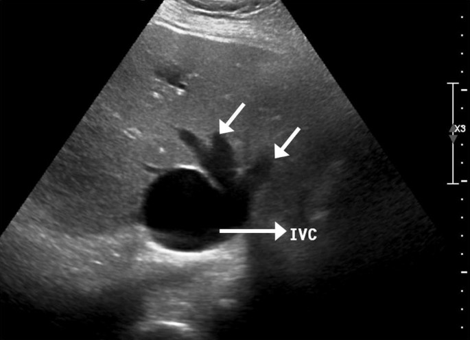


Playboy Bunny Sign Springerlink
Tests to determine the cause of hepatomegaly vary, depending on the suspected cause, but may include Abdominal xray;Hepatomegaly is a term that describes the enlargement of the liver On the other hand, hepatosplenomegaly describes the enlargement of both the liver and the spleen How Liver Size Is Detected Liver size is detected through several methods Imaging methods like ultrasound, magnetic resonance imaging (MRI), and computed tomography (CT) scansAll fetuses were measured for fetal liver length from the top of the right hemidiaphragm to tip of the right liver lobe on coronal image of the fetal abdomen, using highresolution realtime ultrasound with a 2 to 4MHz convex transducer


Hepatomegaly And Diffuse Liver Diseases Springerlink


Fitz Hugh Curtis Syndrome A Diagnosis To Consider In A Woman With Right Upper Quadrant Abdominal Pain Without Gallstones
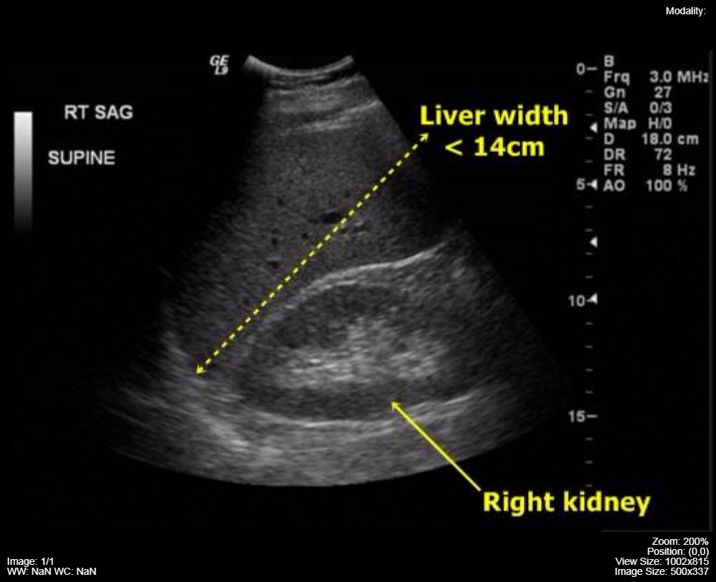
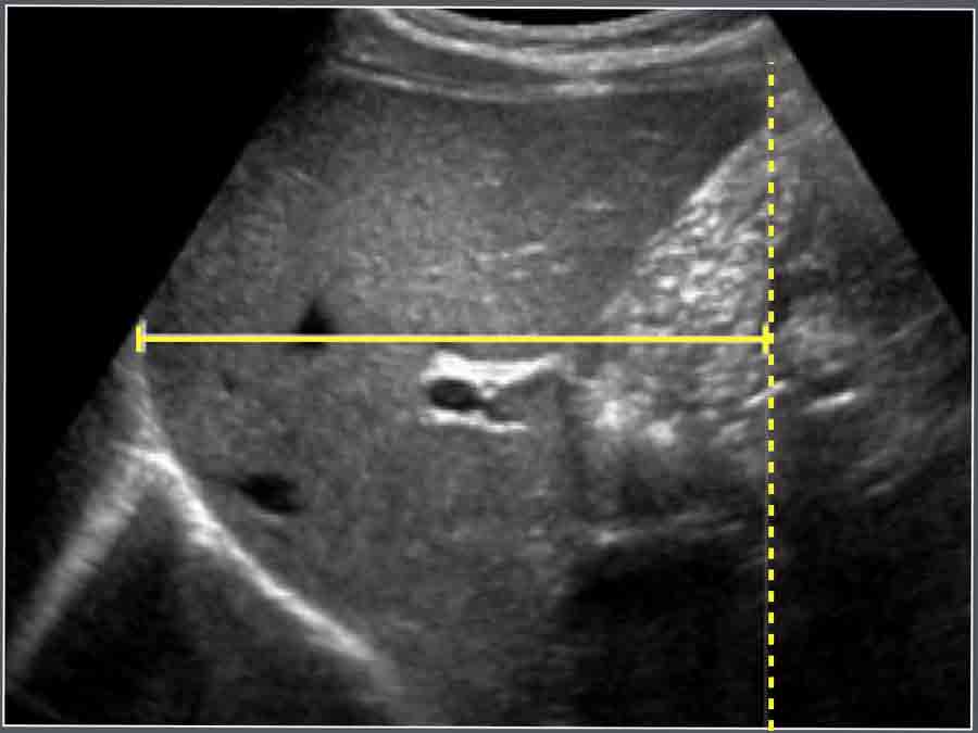
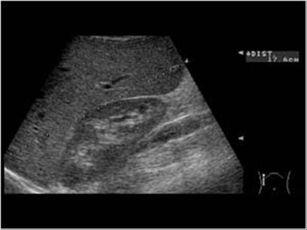
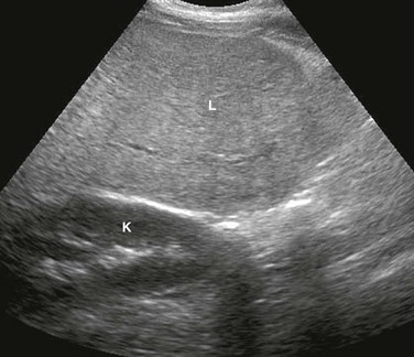
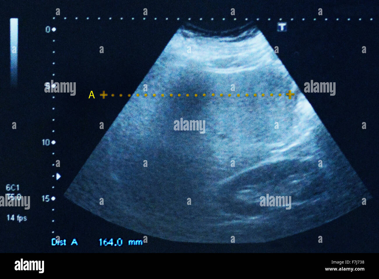
Hepatomegaly • Liver measures more than 15 cm in length (Figure 211) • Commonly seen with infiltrative diseases and masses in the liver Fatty Liver • Mild (early stage) Minimal increase in liver echogenicity Intrahepatic vessels and diaphragm well visualized (Figure 212A) • Moderate (mid stage) Moderate increase in liver echogenicityAssessment of liver size is commonly made on ultrasound or CT, although gross hepatomegaly may be apparent on abdominal radiograph For the adult liver midclavicular line averages cm in craniocaudal length 2 a liver that is longer than cm in the midclavicular line (MCL) is considered enlargedAutopsy determination of hepatomegaly was made using hepatic weight, patient's total body weight, and patient age correlated with pertinent clinical history Results of the autopsy/ultrasound correlation demonstrated that those livers measuring 130 cm or less in the midhepatic line (both supine and left lateral decubitus positions) were normal


Usg Notes Cmud Ultrasound Academy


A Case Of Hepatomegaly The Bmj
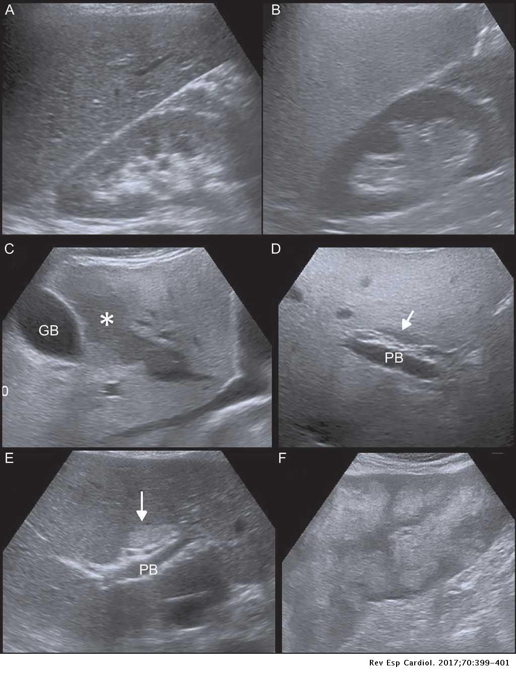
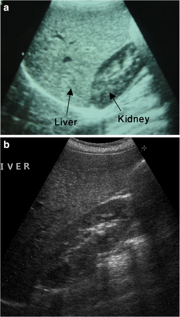
By ultrasound, a normal liver span is usuallyMRI scan of theUltrasound (US) is a commonly used diagnostic tool in the evaluation of the liver and is the most highly recommended imaging modality used in evaluation of suspected liver disease 1 However, as it is largely subjective, characterizing liver echotexture is often difficult for radiologists with limited experience 2


Fetal Hepatomegaly


Pin On Abdominal Pathophysiology Final Exam
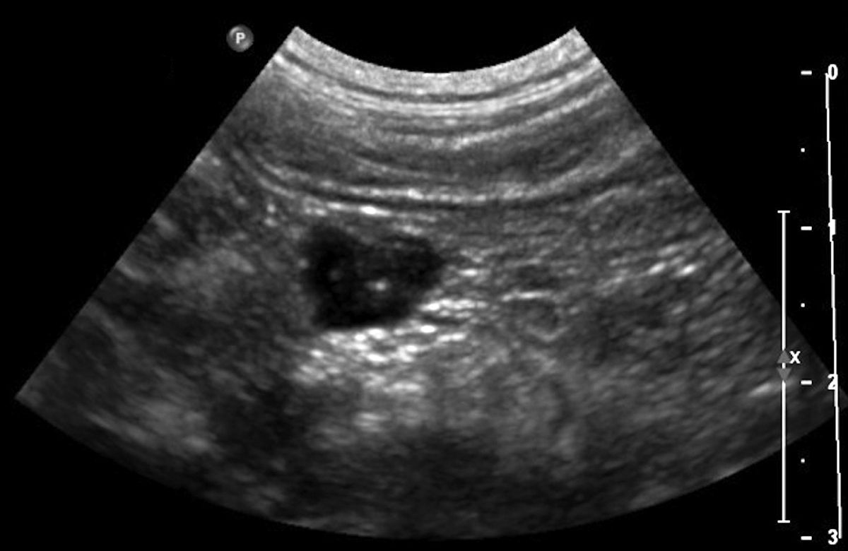
Hepatic ultrasound can be used to determine internal architecture and is better suited than radiography for narrowing the list of considerations for hepatomegaly Visualization of focal hepatomegaly depends on the degree of enlargement and the lobe affected Sonography is an effective, noninvasive, safe, and inexpensive technique for measurement of the liver Measurements of the liver using 2D ultrasound aid in diagnosing and tracking liver disease and in surgical planning Multiple studies have developed techniques to measure the adult liver using 2D ultrasound What Is a Heterogeneous Liver?


Fetal Hepatomegaly Causes And Associations Radiographics


Favorite Tweet Category Monday Poster Session P7 Primary Amyloidosis Presenting As Hepatomegaly In A Patient With Undiagnosed Multiple Myeloma A Challenging Case Of Pneumococcal Meningitis And Hepatomegaly With Obstructive Jaundice
Ultrasound results showed that I Hepatomegaly with severe steatosis I recently had an executive check up and part of the package was ultrasound and checking the sgpt levels The radiologist found on the result that I have "Enlarged with severe increased reflectivity of its parenchyma", the liver size was 163 cm Hepatomegaly is often a sign that the tissue within the liver isn't functioning properly Taking certain medications, such as amiodarone and statins, may also cause liver injuryHepatomegaly, also known as an enlarged liver, isn't considered a disease, but it's usually a sign something is wrong with the liver This video describes th


Hepatomegaly Pelvic Ultrasound Stock Image C027 2735 Science Photo Library
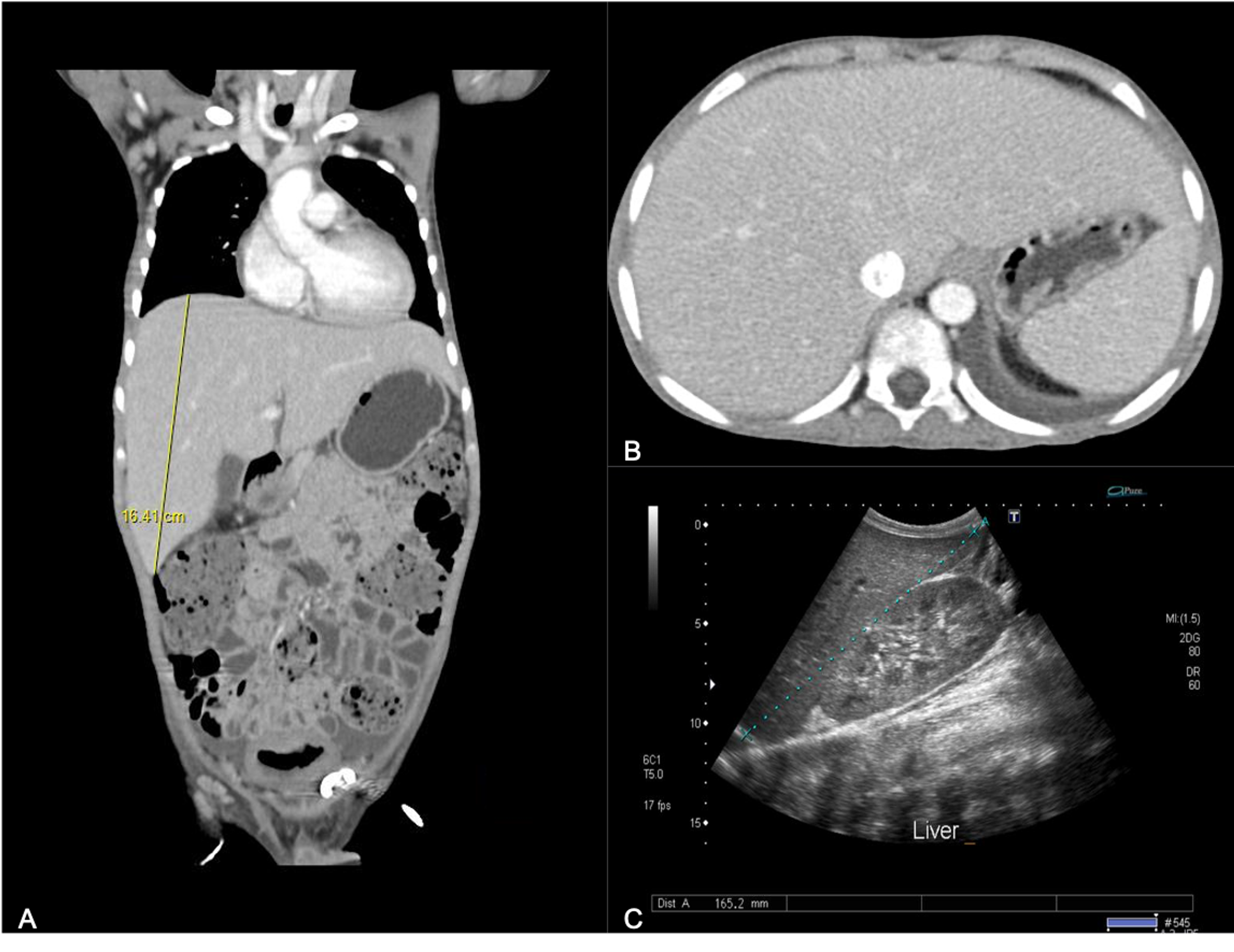


Cureus Mauriac Syndrome Still Exists In Poorly Controlled Type 1 Diabetes A Report Of Two Cases And Literature Review
RUQ ultrasound is useful in assessing three common problems Right upper quadrant and epigastric pain Jaundice Ascites There are four common practical applications for bedside ultrasound of the RUQ, including the detection of Gallstones Cholecystitis Dilated bile ducts and biliary obstruction AscitesLiver function tests, including blood clotting tests;By ultrasound, a normal liver span is usually


Animal Liver Disease Long Beach Animal Hospital
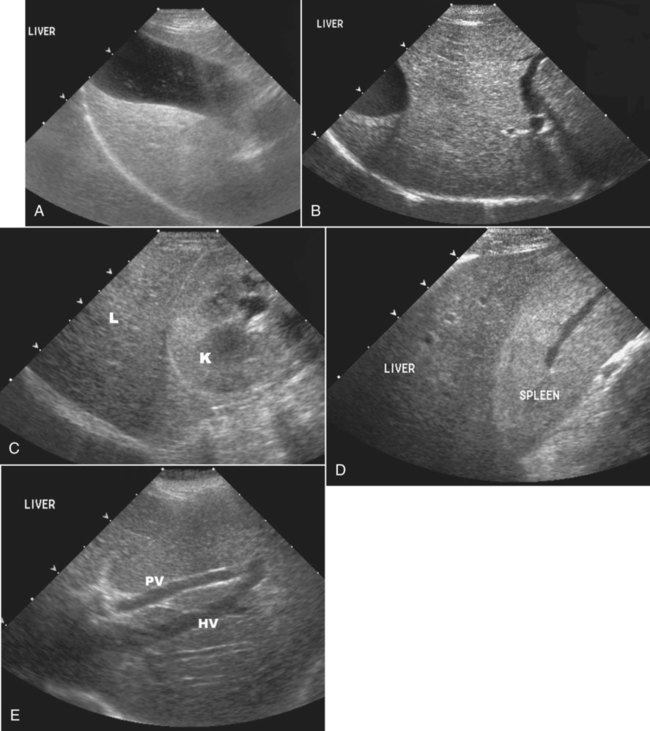


The Liver And Spleen Veterian Key
Diagnosed with "borderline hepatomegaly with mild fatty infiltration of liver and pancreas" in ultrasound please suggest the reasons and treatment? R160 is a billable/specific ICD10CM code that can be used to indicate a diagnosis for reimbursement purposes The 21 edition of ICD10CM R160 became effective on This is the American ICD10CM version of R160 other international versions of ICD10 R160 may differ Applicable To The initial strategy in cirrhosis should be the measurement of αfetoprotein (AFP) followed by ultrasound, contrast CT or magnetic resonance imaging Primary hepatic lymphoma is rare and can present as solitary or multiple masses, as a diffuse hepatic involvement with hepatomegaly, or as hepatic failure with elevated LDH
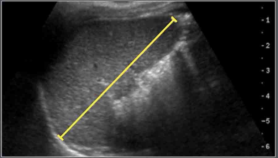


The Radiology Assistant Normal Values Ultrasound


Right Lobe Of Liver An Overview Sciencedirect Topics
Hepatomegaly in dogs is typically recognized during presentation for nonspecific signs such as weakness, lethargy and inappetence These patients are sometimes jaundiced Causes infectious and toxic causes are typically associated with younger animals while neoplastic and cardiac causes are seen more frequently in older dogs Hepatomegaly is a term used to describe a liver that is enlarged beyond its normal dimensions, and ultrasound is often a front line investigation in the suspicion of hepatomegaly This study sought to develop a reference range for the size of the normal liver using a simple, reliable and valid measurement technique Ultrasound findings in the study population were as follows 572% steatosis, 495% hepatomegaly, 17% heterogeneous liver, and 16% portal hypertension Of note, 77% had at least one ultrasound abnormality, and 45% had ≥2


Figure1 A B Ultrasound Images Showing Severe Hepatomegaly And Fatty Download Scientific Diagram


Imaging Of The Liver And Pancreas Vet Focus
Dr Mark Hoepfner answered General Surgery 39 years experience Varies An enlarged fatty liver ( hepatomegaly) can be seen on ultrasound This may be a more common condition in diabetics or those that are obese or overweight Fat cells build up in the liver, and in some cases liver damage can occur Hepatosplenomegaly (HPM) is a disorder where both the liver and spleen swell beyond their normal size, due to one of a number of causes The name of this condition — hepatosplenomegaly — comes


Hepatomegaly In Cats Vetlexicon Felis From Vetlexicon Definitive Veterinary Intelligence


Journals Sagepub Com Doi Pdf 10 1177


Hepatomegaly Wikipedia


Favorite Tweet Category Monday Poster Session P7 Primary Amyloidosis Presenting As Hepatomegaly In A Patient With Undiagnosed Multiple Myeloma A Challenging Case Of Pneumococcal Meningitis And Hepatomegaly With Obstructive Jaundice


Q Tbn And9gctgtu0dlmgmgagdl75oobp8bgvzztksormyeuozizq Usqp Cau
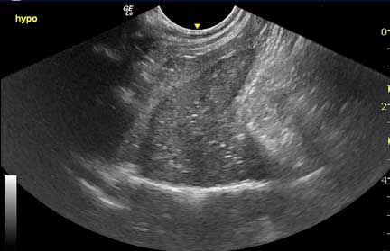


Animal Liver Disease Long Beach Animal Hospital


Fetal Hepatomegaly Causes And Associations Radiographics


Doc Does My Pet Really Need All These


Hepatomegaly Enlarged Liver Symptoms Causes And Treatment


Fetal Hepatomegaly Causes And Associations Radiographics


What Is The Differential Diagnosis Of Hepatomegaly Pediatriceducation Org


Role Of Abdominal Ultrasound In The Diagnosis Of Typhoid Fever In Pediatric Patients Sciencedirect


Abdominal Ultrasonography Showing Hepatomegaly With Increased Hepatic Download Scientific Diagram


Abd Ultrasound Liver Pathology Flashcards Quizlet


Ultrasound Of Enlarged Liver Download Scientific Diagram
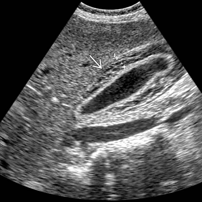


Viral Hepatitis Radiology Key


View Image
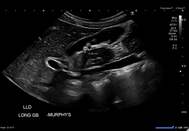


Hereditary Spherocytosis Radiology Case Radiopaedia Org


Ultrasound Us Undergraduate Diagnostic Imaging Fundamentals


Figure 1 Artificial Neural Network Application In The Diagnosis Of Disease Conditions With Liver Ultrasound Images


The Radiology Assistant Normal Values Ultrasound


Chronic Liver Disease Among Adult Patients With Sickle Cell Anemia In Steady State In Ile Ife Nigeria Oguntoye Journal Of Gastroenterology And Hepatology Research


Liver And Cardiovascular Disease What Cardiologists Need To Know About Ultrasound Findings Revista Espanola De Cardiologia


Imaging Of A Rare Location Extra Nodal Lymphoma Lesions Eurorad


Hepatomegaly Hoopla Ultrasound Tips Tricks


Imaging Of The Liver And Pancreas Vet Focus


Anatomical Criteria To Measure The Adult Right Liver Lobe By Ultrasound Abstract Europe Pmc


Ultrasound In Acute Viral Hepatitis Does It Have Any Role Maurya V Ravikumar R Gopinath M Ram B Med J Dy Patil Vidyapeeth


Prevalence Of Hepatopathy In Type 1 Diabetic Children Bmc Pediatrics Full Text


Normal Liver Ultrasound How To


Abdomen And Retroperitoneum 1 4 Spleen Case 1 4 5 Malignant Lesions Of The Spleen Ultrasound Cases


Parenchymal Liver Disease Radiology Key


Dedicated To The Mission Of Bringing Free Or Low Cost Educational Materials And Information To The Global Ultrasound Community


Hepatomegaly Ultrasound Stock Photo Alamy


Small Animal Abdominal Ultrasonography Part 2 Liver Gallbladder Today S Veterinary Practice


Abdominal Ultrasound There Is Mild Hepatomegaly With Increased Liver Download Scientific Diagram


Ultrasound Us Undergraduate Diagnostic Imaging Fundamentals


The Radiology Assistant Normal Values Ultrasound


Ultrasound In The Assessment Of Hepatomegaly A Simple Technique To Determine An Enlarged Liver Using Reliable And Valid Measurements Childs 16 Sonography Wiley Online Library


3


The Liver Part 1 Normal Appearance Imv Imaging


Enlarged Liver Riedel Lobe Radiology Case Radiopaedia Org


1


Hepatomegaly Ultrasound Stock Image C026 9499 Science Photo Library
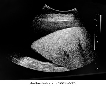


Hepatomegaly Images Stock Photos Vectors Shutterstock


Liver 3 Times Normal Size Novocom Top


Hepatomegaly Hoopla Ultrasound Tips Tricks


Factors Affecting Liver Size Kratzer 03 Journal Of Ultrasound In Medicine Wiley Online Library


Epos
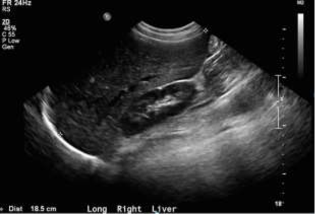


Riedel Fight Song A Case Report Of Riedel Lobe Presumed To Be Hepatomegaly Semantic Scholar


Ultrasound Of Riedel Lobe Celus


Q Tbn And9gctjckyy0t1y4un0vmublxeomddhj7bnr5v6awiefj54y0ohszr Usqp Cau


Hepatomegaly Wikipedia


Ultrasound Video Showing Hepatomegaly With Liver Approaching Kissing The Spleen Youtube


Hepatomegaly In Dogs Vetlexicon Canis From Vetlexicon Definitive Veterinary Intelligence
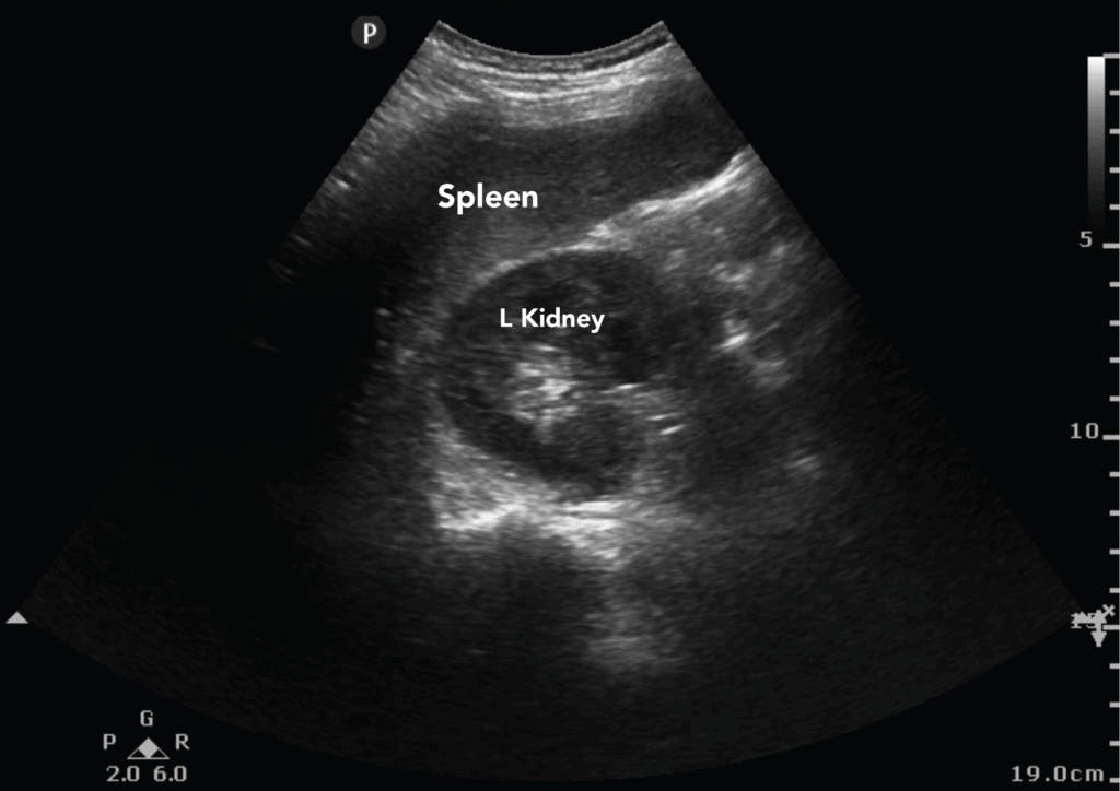


Abdominal Ultrasound Made Easy Step By Step Guide Pocus 101


Ultrasound In The Assessment Of Hepatomegaly A Simple Technique To Determine An Enlarged Liver Using Reliable And Valid Measurements Childs 16 Sonography Wiley Online Library
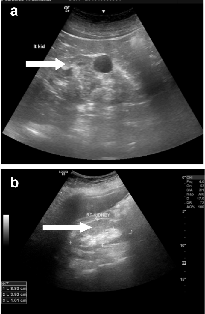


Diagnostic Value Of Abdominal Sonography In Confirmed Covid 19 Intensive Care Patients Egyptian Journal Of Radiology And Nuclear Medicine Full Text


Sir My Baby Girl Has Severe Stomach Pain In Last Week Doctor Suggested For A Ultrasound In Ultrasound She Diagnosed With Mild Hepatomegaly But The Doctor Said That Is Not The Reason Behind Stomach
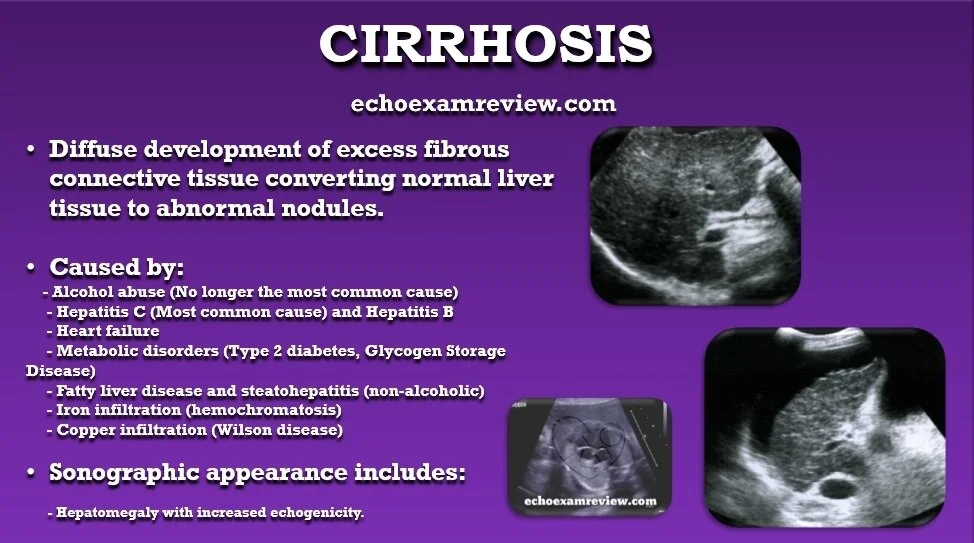


Ultrasound Board Review


Longitudinal Sonogram Of Liver Showing Hepatomegaly And Increased Download Scientific Diagram
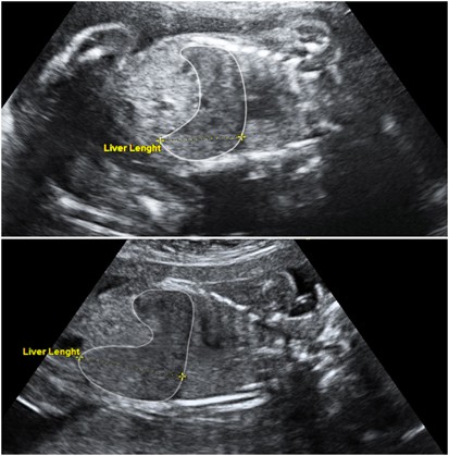


Fetal Liver Length Measurement At Mid Pregnancy Among Fetuses At Risk As A Predictor Of Hemoglobin Bart S Disease Journal Of Perinatology


Journals Sagepub Com Doi Pdf 10 1177
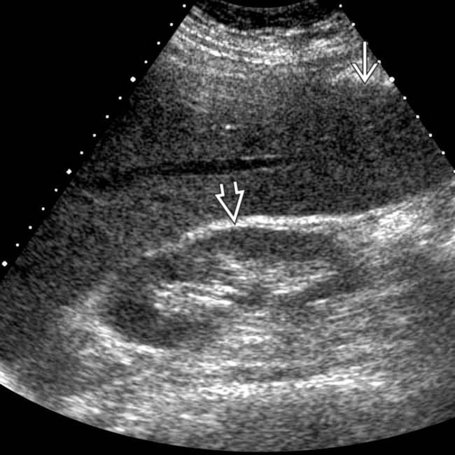


Hepatomegaly Clinical Gate


Hemochromatosis Rare Characterized By Excess Iron Deposits Throughout Body May Cause Cirrhosis Sonographic Findings Hepat Sonography Gallbladder Cirrhosis


The Sonographic Dimensions Of The Liver At Normal Subjects Compared To Patients With Malaria Science Publishing Group


Figure 1 From Noninvasive Assessment Of Liver Steatosis Using Ultrasound Methods Semantic Scholar


Gray Scale Ultrasound Image Of The Liver Revealed Features Of Download Scientific Diagram


Journal Of Aids And Hiv Research Correlation Of Hepatobiliary Ultrasonographic Findings With Cd4cell Count And Liver Enzymes In Adult Hiv Aids Patients In Jos


Fetal Hepatomegaly Causes And Associations Radiographics


Prevalence Of Sonographic Signs In Children With Acute Hepatitis In Zahedan City Southeast Of Iran International Journal Of Infection Full Text
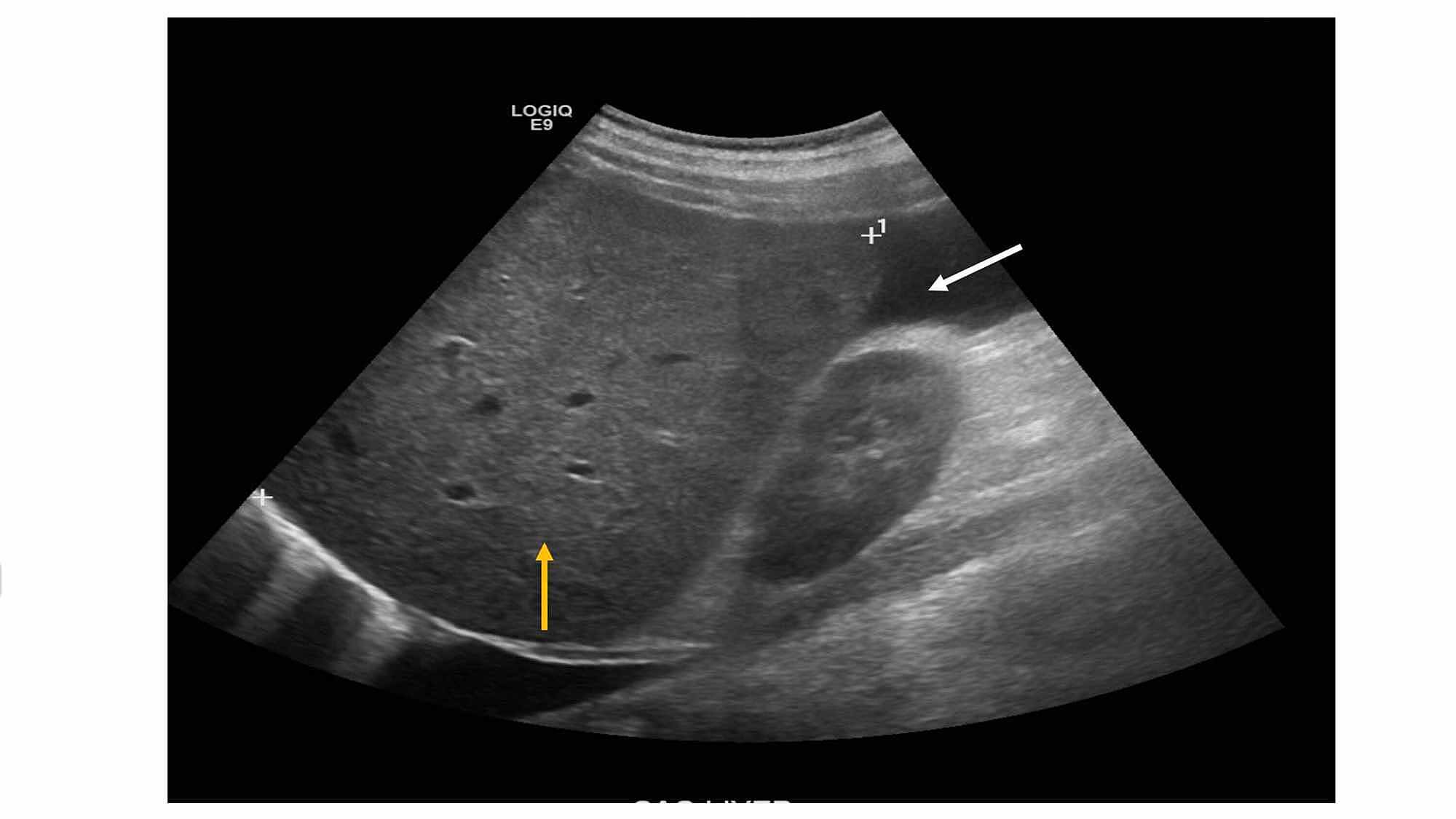


Cureus Amyloidosis Masquerading As Alcohol Related Cirrhosis


Liver Ultrasound Abnormalities In Alcohol Use Disorder Intechopen


Acep American College Of Emergency Physicians


Liver Pamesono
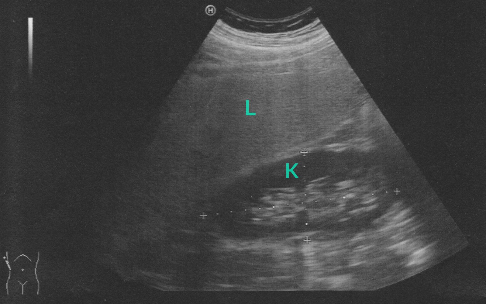


Alcoholic Liver Disease Amboss
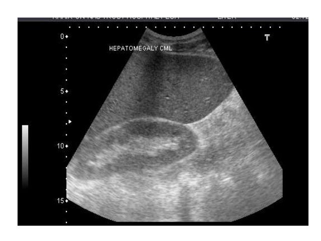


Presentation1 Liver Ultrasound
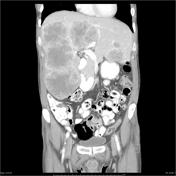


Hepatomegaly Radiology Reference Article Radiopaedia Org


Normal Liver Ultrasound How To


Liver And Cardiovascular Disease What Cardiologists Need To Know About Ultrasound Findings Revista Espanola De Cardiologia


How Severe Is Hepatomegaly With Fatty Liver Grade 2 Quora
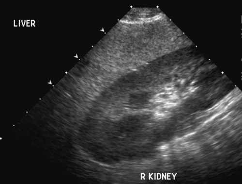


The Liver And Spleen Veterian Key


コメント
コメントを投稿